That’s “not safe for work”, not “national science foundation” up there in the acronym. It was surely inevitable that this amazing, subtle and elegant technology would eventually be applied to more scatological pursuits. The following paper is a classic in this genre.
Magnetic resonance imaging of male and female genitals during coitus and female sexual arousal.
OBJECTIVE: To find out whether taking images of the male and female genitals during coitus is feasible and to find out whether former and current ideas about the anatomy during sexual intercourse and during female sexual arousal are based on assumptions or on facts. DESIGN: Observational study. SETTING: University hospital in the Netherlands. METHODS: Magnetic resonance imaging was used to study the female sexual response and the male and female genitals during coitus. Thirteen experiments were performed with eight couples and three single women. RESULTS: The images obtained showed that during intercourse in the “missionary position” the penis has the shape of a boomerang and 1/3 of its length consists of the root of the penis. During female sexual arousal without intercourse the uterus was raised and the anterior vaginal wall lengthened. The size of the uterus did not increase during sexual arousal. CONCLUSION: Taking magnetic resonance images of the male and female genitals during coitus is feasible and contributes to understanding of anatomy.
Schultz et al, BMJ. 1999 Dec 18-25;319(7225):1596-600. PMID 10600954.
To be absolutely fair, this was done by members of the Department of Gynaecology at the University Hospital in Groningen (Netherlands, of course!). So this wasn’t a spurious application. The methods section describes the basic setup:
The tube in which the couple would have intercourse stood in a room next to a control room where the searchers were sitting behind the scanning console and screen. An improvised curtain covered the window between the two rooms, so the intercom was the only means of communication. Imaging was first done in a 1.5 Tesla Philips magnet system (Gyroscan S15) and later in a 1.5 Tesla magnet system from Siemens Vision. To increase the space in the tube, the table was removed: the internal diameter of the tube is then 50 cm. The participants were asked to lie with pelvises near the marked centre of the tube and not to move during imaging. After a preview, 10 mm thick sagittal images were taken with a half-Fourier acquisition single shot turbo SE T2 weighted pulse sequence (HASTE). The echo time was 64 ms, with a repetition time of 4.4 ms. With this fast acquisition technique, 11 slices of relatively good quality were obtained within 14 seconds.
The volunteers were shown the equipment in the two rooms, and personal and gynaecological histories were taken. The experimental procedure was explained, and all investigators left the imaging room. After a preliminary image for positioning the true pelvis of the woman was taken, the first image was taken with her lying on her back (image 1). Then the male was asked to climb into the tube and begin face to face coitus in the superior position (image 2). After this shot—successful or not—the man was asked to leave the tube and the woman was asked to stimulate her clitoris manually and to inform the researchers by intercom when she had reached the preorgasmic stage. Then she stopped the autostimulation for a third image (image 3). After that image was taken the woman restarted the stimulation to achieve an orgasm. Twenty minutes after the orgasm, the fourth image was taken (image 4). At the end of the experiment, the images were evaluated in the presence of the participants.
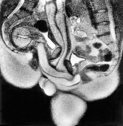
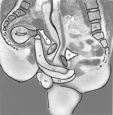
Naturally, HASTE would be the most appropriate pulse sequence. The results show more internal contortion than one might expect, but were not otherwise particularly surprising. Here’s the, ahem, money shot:


caption: Midsagittal image of the anatomy of sexual intercourse (experiment 12). P=penis, Ur=urethra, Pe=perineum, U=uterus, S=symphysis, B=bladder, I=intestine, L5=lumbar 5, Sc=scrotum
In the Discussion section, the authors also get the unique honor of proving that Leonardo da Vinci underestimated the size of the male organ. Take that, Leonardo! The paper concludes,
What started as artistic and scientific curiosity has now been realised. We have shown that magnetic resonance images of the female sexual response and the male and female genitals during coitus are feasible and beautiful; that the penis during intercourse in the “missionary position” has the shape of a boomerang and not of an S as drawn by Dickinson; and that, in contrast to the findings of Masters and Johnson, there was no evidence of an increase in the volume of the uterus during sexual arousal.
It should be noted that PZ Meyers talked about this paper last April, even though it’s not exactly up his area of expertise. The subject matter certainly lends itself to interdisciplinary collaboration!
One response to “NSFW MRI: Sex at 1.5T”
[…] a not work safe post on […]